|
Histology
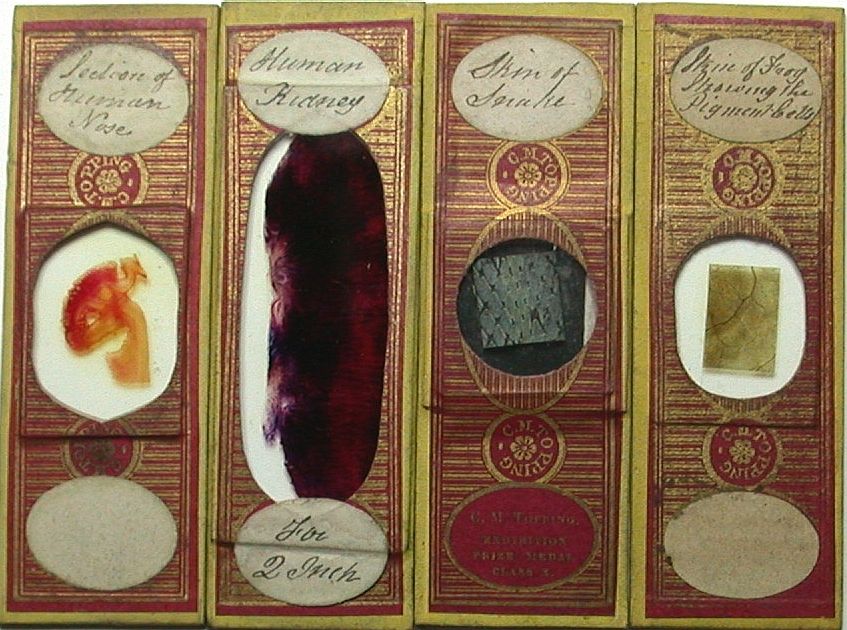
mounts are often what comes to mind for many
people when "old microscope slides" are mentioned. Without
doubt,
our
knowledge of the human body's structures and processes, as well of
those of animals and plants, owes a great deal to the early slide
makers of the Victorian era. Before about 1850, most mounted histology
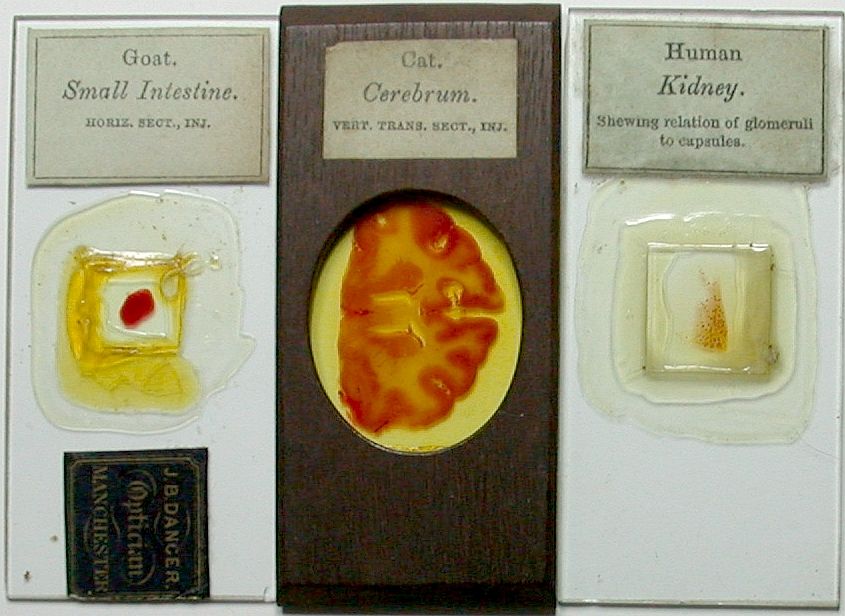
specimens were thick sections of tissue, sometimes with the vascular
structures injected with
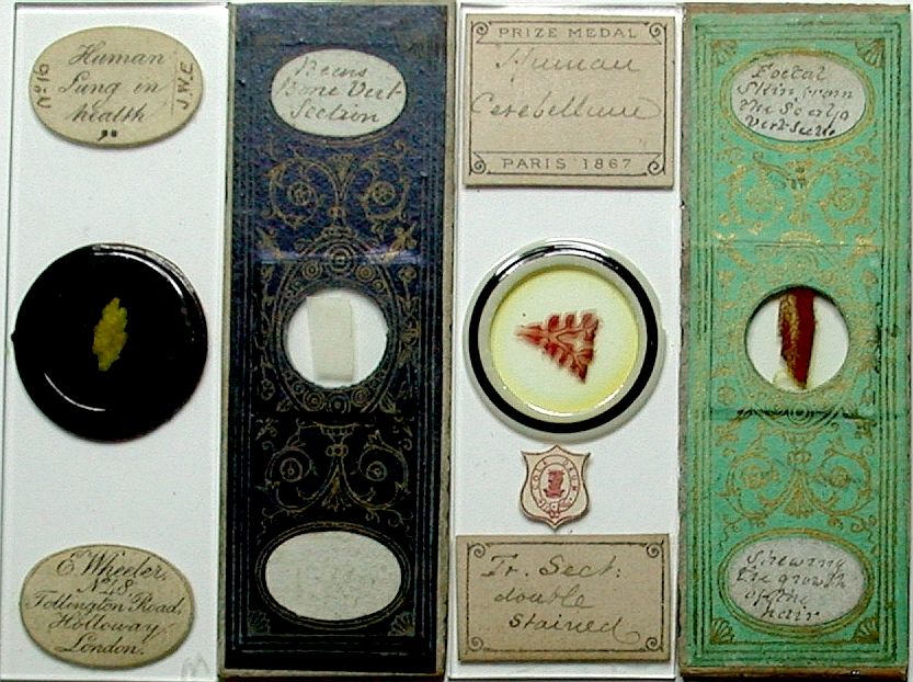
various coloured substances to make them more visible, and usually
mounted as "deep cell" fluid mounts (see top row, 2nd from
left - engraved "Skin from the Ear of the Cat showing the
Vessels of the bulbs, & the hairs Hett, 1849"
). These thick preparations could only be examined microscopically
with reflected (incident) top lighting, and provided limited
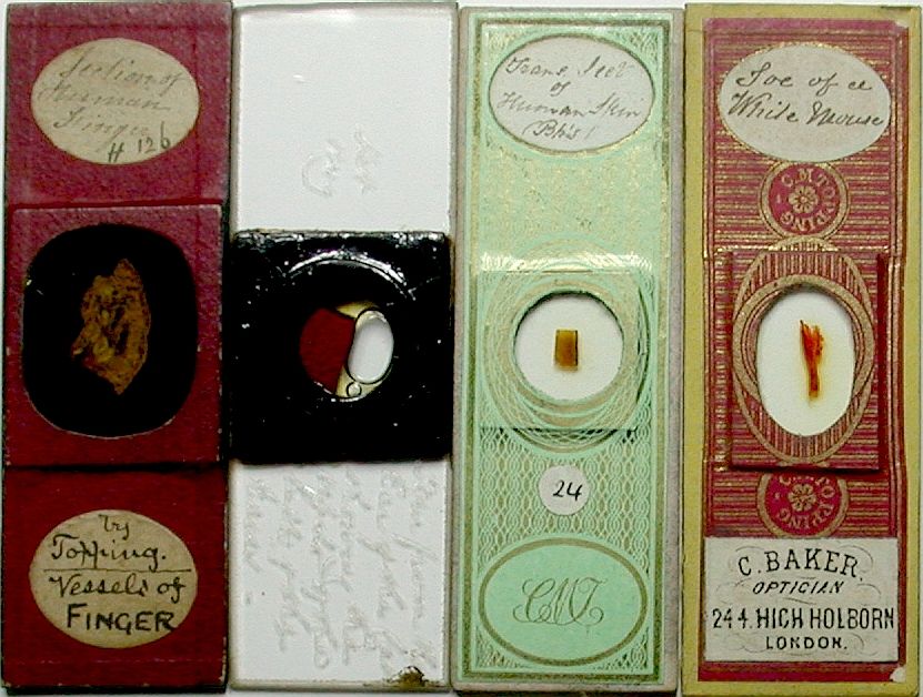
information. The early commercial
slide preparers such as C. M. Topping, J. T. Norman, A. C. Cole, and
E. Wheeler often worked closely with leading
medical professionals of the era to further current research and knowledge in
the fields of medicine and surgery. These individuals, by their
dedicated efforts, helped develop the techniques in preparation and
preservation that allowed the research underlying the remarkable advances in medicine that
we benefit from today. An important early development was consistent
thin sectioning of tissue specimens, and preparation/preservation
techniques that enabled use of
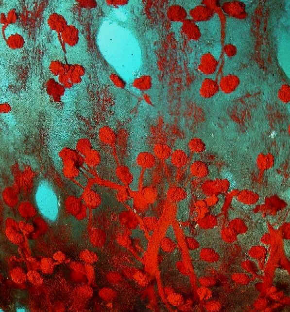
transmitted (through the specimen) lighting. This advance allowed
microscopic examination of the minute structures, helping open the door to
a better understanding of the processes of disease, and the development of new
effective treatments. A wide variety of histology mounts were
prepared by most of the more capable preparers, covering all the
major tissue types (both healthy and diseased) of both human and
animal species. |